

A craniopharyngioma is a tumor that grows near the pituitary gland in the brain. These tumors tend to occur in children more frequently than adults. Craniopharyngiomas frequently contain both solid and cystic parts that usually enlarge over time to compress the neighboring structures in the brain such as the optic nerves, the pituitary gland, and even the ventricular system.
What Causes a Craniopharyngioma?
The exact cause of craniopharyngiomas is not clear. The prevailing belief is that these tumors arise from a cluster of cells that assumes an abnormal location near the pituitary gland during fetal development.
How Are Craniopharyngiomas Discovered?
Craniopharyngiomas compress neighboring structures in the brain as they grow. They can impair vision by compressing the optic nerves and they can cause a host of hormonal abnormalities by compressing the pituitary gland. They can even result in hydrocephalus (the build-up of cerebrospinal fluid in the brain) by compressing the ventricular system leading to lethargy, headache, nausea, and vomiting.
The diagnosis of a craniopharyngioma is often confirmed by obtaining a CT scan and/or an MRI of the brain. In addition to imaging studies of the brain, a thorough endocrine and opthomological evaluation is usually obtained.
How Are Craniopharyngiomas Treated?
Surgical resection is the mainstay of treatment for craniopharyngiomas. Unfortunately, these tumors tend to grow back if not completely removed. Furthermore, not all tumors are amenable to complete resection as they may be very adherent to neighboring structures. Radiation may also be used, either alone or in addition to surgery. Radiation, however, can injure the pituitary and hypothalamus with resultant adverse effects on hormone production and child development.
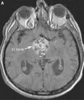
A) Pre-operative axial T1 weighted MRI of the brain with contrast demonstrating the enhancing craniopharyngioma located in the third ventricle.
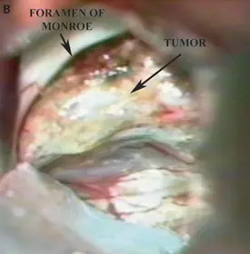
B) Intra-operative photograph showing the craniopharyngioma as it protrudes through the foramen of monroe into the lateral ventricle.
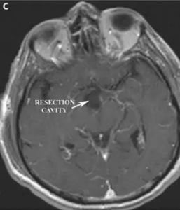
C) Post-operative axial MRI of the brain with contrast demonstrating complete resection of the craniopharyngioma.