


A cavernoma is a small cluster of thin and thick walled venous channels without any intervening neural tissue. A cavernoma may be found near a large vein that drains a large region of brain called a developmental venous anomaly (DVA). A cavernomas, however, lacks and direct connection with any feeding arteries or draining veins. Cavernomas are usually found in the brain and brain stem, rarely are they found in the spinal cord. In 50 percent of case a cavernomas are multiple.
Genetics has been shown to influence cavernoma formation. In fact entire families that have cavernous malformations have been identified and studied. Mutations in several genes have been identified (CCM1, CCM2 and CCM3) that are responsible for the autosomal dominant inheritance patterns of cavernoma in these families. This autosomal dominant inheritance pattern accounts for less than half of cavernomas, and the remaining patients have a spontaneous mutations that lead to the formation of these cavernomas. Radiation treatments to the brain have been associated with the spontaneous formation of cavernomas, particularly if the radiation was administered in childhood.
Patients with cavernomas may present with new onset seizures, progressive neurological deficit and occasionally cerebral hemorrhages. Cavernomas tend to have repeated small hemorrhages that are rarely neurologically devastating. The annual risk of a cavernoma bleeding ranges from 0.2 to 2 percent. Cavenomas are usually diagnosed on MRI as they have a very characteristic appearance.
The treatment strategy for a cavernoma depends on its location in the brain and spinal cord. Cavernomas that are in surgically accessible regions of the brain usually can be removed with a reasonable degree of safely. If a cavernoma is located in the brain stem or spinal cord surgical excision carries a greater risk of post-operative neurological deficit. For this reason, brain and spinal cavernomas are frequently observed and surgical resection is reserved for patients who have repeated hemorrhages associated with neurological deterioration. Surgery is the only available treatment for cavernomas and it is well documented that they do not respond to radiation therapy. Some families of patients with multiple cavernomas and/or multiple family members with cavernomas may benefit from genetic counseling.
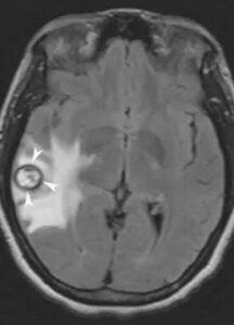
Axial flair sequence of the brain demonstrating a right temporal cavernoma. Note the dark ring (arrowheads) around the cavernoma. This ring, called a hemosiderin ring, arises from old blood products that are deposited around the perimeter of the cavernoma following small hemorrhagic events. This hemosiderin ring is characteristic of cavernomas.