

What Causes Lumbar Spinal Stenosis?
For most patients, lumbar stenosis is an acquired disease process brought about by chronic wear and tear on the spine. Chronic wear and tear leads to a host of degenerative changes that result in a progressively narrow spinal canal. Boney growths along the facet joints of the spine along with thickening of the spinal ligaments and disc herniations all contribute to narrowing the spinal canal. Genetics, however, is believed to contribute to the development of spinal stenosis in some patients. Just as some people are tall and others are short, some people have very large spinal canals and others have very small ones. Achondroplastic dwarves, for example, have a genetic abnormality that affects the growth and development of their bones and for this reason they are more likely to develop symptomatic spinal stenosis.
How Is Lumbar Spinal Stenosis Diagnosed?
The classic symptoms of lumbar stenosis include pain in the buttocks, legs, and feet brought about by walking that is relieved with rest or postural changes such as sitting or bending forward. Not all patients complain of pain, some complain of leg cramping and still others note leg weakness or fatigue. These symptoms, formally known as neurogenic claudication, may be unilateral or bilateral.
In some patients neurogenic claudication may be difficult to differentiate from claudication symptoms due to vascular disease in the lower extremities. Leg pain from vascular claudication, unlike neurogenic claudication, may persist at rest and is associated with the following findings: pulselessness, paraesthesias, paralysis, pallor and pain in one or both extremities. In cases where vascular claudication is suspected evaluation by a vascular specialist may be required.
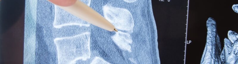
Once neurogenic claudication is suspected an MRI or CT scan of the lumbar spine demonstrates the severity and location of the spinal stenosis. The information provided by these studies is critical for planning an effective treatment.
How Is Lumbar Spinal Stenosis Treated?
Surgical decompression of the nerve roots is the mainstay in the treatment for lumbar stenosis. A lumbar laminectomy is the most common form or surgical treatment and involves removing the bone and ligaments (spinous process, medial facet joint, and ligamnetum flavum) from the posterior aspect of the spinal canal. Removal of this bone and ligament effectively increases the size of the spinal canal and relieves the compression of the nerve roots. In some cases the patient may have underlying spinal instability that requiring instrumented stabilization and fusion.
Certain minimally invasive techniques have recently evolved to treat lumbar stenosis. A technique of interspinous distraction using an implanted device called the X STOP has proven effective in treating patients with single level lumbar stenosis. The X STOP is inserted in between the spinous processes of the lumbar spine increasing the separation between them. By distracting the spinous processes in this way, the X STOP is able to increase the diameter of the spinal canal alleviating the symptoms of spinal stenosis. The X STOP also increases the size of the opening in the lumbar spine where the nerve roots exit the spinal canal (neural foramen). The X STOP is best suited for patients who have leg pain rather than back pain, and who experience some improvement in their symptoms when they bend forward.
Patients who either are not healthy enough for surgery or do not wish to undergo surgery may be candidates for epidural steroid injections. Epidural steroid injections may provide temporary relief of symptoms. During an epidural steroid injection a needle is passed through the skin into the spinal canal using x-ray guidance. Once in the proper location a mixture of steroid (a strong anti-inflammatory medication) and local anesthetic is administered.
Physical therapy alone is an alternative method of treatment. This, however, results in symptomatic relief in very few patients.