What Causes Cervical Spinal Stenosis?
For most patients, cervical stenosis is an acquired disease process brought about by chronic wear and tear on the spine. Chronic wear and tear leads to a host of degenerative changes that result in a progressively narrow spinal canal. Boney growths along the facet joints of the spine along with thickening of the spinal ligaments and disc herniations all contribute to narrowing the spinal canal. Genetics, however, is believed to contribute to the development of spinal stenosis in some patients. Just as some people are tall and others are short, some people have very large spinal canals and others have very small ones. Achondroplastic dwarves, for example, have a genetic abnormality that affects the growth and development of their bones and for this reason they are more likely to develop symptomatic spinal stenosis.
How is Cervical Spinal Stenosis Diagnosed?
Patients with stenosis of the cervical spine experience symptoms from chronic compression of the spinal cord. These patients often complain of numbness and tingling in the hands as well as progressive difficulty using their hands to perform fine tasks such as writing their name with a pen or buttoning their shirt. Patients with cervical spinal stenosis may also experience trouble walking and frequent falls. Weakness in the hip flexor muscles (iliopsoas muscles) is also a feature of cervical stenosis. In some patients these findings, known collectively as myelopathic symptoms, are mild resulting in minor impairment. Other patients with advanced forms of cervical myelopathy may be bed bound due to severe arm and leg weakness.
Once cervical stenosis is suspected an MRI of the cervical spine helps delineate the location and severity of the compression of the cervical spinal cord. A CT scan of the cervical spine provides additional information about the boney anatomy of the cervical spine, an important consideration in planning treatment.
How is Cervical Spinal Stenosis Treated?
Surgery is the mainstay in the treatment of symptomatic cervical stenosis. Not all patients with symptomatic cervical stenosis require an operation. For example, patients who have mild symptoms may be observed. Surgical intervention is typically reserved for patients with progressive symptoms, patients with weakness of the arms and legs, or patients with severe numbness or pain.
The surgical treatment of cervical stenosis depends upon the extent and location of the compression of the cervical spinal cord. Each case is assessed on an individual basis to determine the optimal surgical approach. Surgery often involves decompression of the spinal cord through an anterior approach, a posterior approach, or a combination of the two. In general, anterior approaches to the cervical spine are often used when the compression of the spinal cord arises from pathology arising from the disc spaces and vertebral bodies. Posterior approaches to the cervical spine are used when the compression is from the ligaments, lamina, and facet joints that comprise the back of the spine.
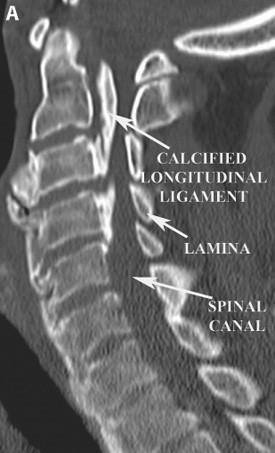
A) A sagittal CT scan of the cervical spine demonstrating severe narrowing of the spinal canal due to calcification of the posterior longitudinal ligament.Note that bones that make up the back of the spinal canal are called the lamina.
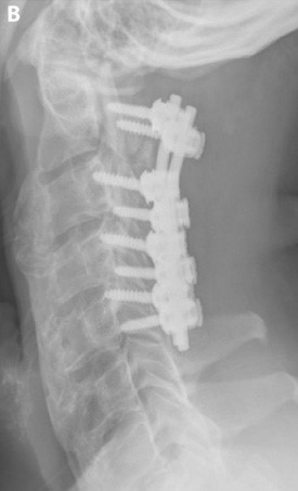
B) Post-operative lateral cervical spine x-ray demonstrating a laminectomy (removal of the bones that make up the posterior portion of the spinal canal) and instrumented stabilization of the cervical spine with lateral mass screws and rods. The laminectomy effectively increases the size of the spinal canal.